 Leanpub
In both the particle-based and automaton model for Turing patterns, we observed that the model is fine-tuned, meaning that very slight changes in parameter values can lead to significant changes in the system. These changes could convert spots to stripes, or they could influence how clearly defined the boundaries of the Turing patterns are.
The figure below shows how the Turing patterns produced by the Gray-Scott model change as the kill and feed rates vary. The square at position (x, y) shows the pattern obtained as the result of a Gray-Scott simulation with kill rate x and feed rate y. Notice how much the patterns change! You may like to tweak the parameters of the Gray-Scott simulation from the previous lesson to see if you can reproduce these differing patterns.
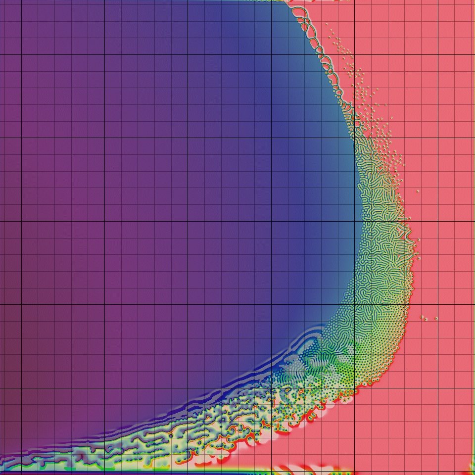 Changing kill (x-axis) and feed (y-axis) parameters greatly affects the Turing patterns obtained in the Gray-Scott model. Each small square shows the patterns obtained from a given choice of feed and kill rate. Note that many choices of parameters do not produce Turing patterns, which only result from a narrow “sweet spot” band of parameter choices. Image courtesy: Robert Munafo.1
Later in this course, we will see an example of a biological system that is the opposite of fine-tuned. In a robust system, perturbations such as parameter variations do not lead to substantive changes in the ultimate behavior of the system. Robustness is vital for processes, like your heartbeat, that must be resilient to small environmental changes.
It turns out that although Turing’s work offers a compelling argument for how zebras might have gotten their stripes, the cellular mechanism causing these stripes to form remains unidentified. However, researchers have shown that the skin of zebrafish does exhibit Turing patterns because two types of pigment cells serve as “particles” following a reaction-diffusion model much like the one we presented in this prologue.2
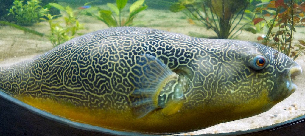
Finally, take a look at the following two photos of giant pufferfish.34 Genetically, these fish are practically identical, but their skin patterns are very different. What may seem like a drastic change from spots to stripes is likely attributable to a small change of parameters in a fine-tuned biological system that, like much of life, is powered by randomness.

A final note
Thank you for making it this far! We hope that you are enjoying the course. You can visit the exercises for this module or skip ahead to the next module by clicking on the appropriate button below. We also ask that you complete the course survey if you have not done so already.
-
“Reaction-Diffusion by the Gray-Scott Model: Pearson’s Parametrization” © 1996-2020 Robert P. Munafo https://mrob.com/pub/comp/xmorphia/index.html ↩
-
Nakamasu, A., Takahashi, G., Kanbe, A., & Kondo, S. (2009). Interactions between zebrafish pigment cells responsible for the generation of Turing patterns. Proceedings of the National Academy of Sciences of the United States of America, 106(21), 8429–8434. https://doi.org/10.1073/pnas.0808622106 ↩
-
NSG Coghlan, 2006 Creative Commons Attribution-Share Alike 3.0 Unported ↩
-
Chiswick Chap, 20 February 2012, Creative Commons Attribution-Share Alike 3.0 Unported ↩
